Hip Thrust And Back Squat Training Produce Comparable Increases In Gluteal Muscle Size And Exhibit Similar Transfer To The Deadlift In Untrained Individuals!
A remarkable group of researchers collaborated to carry out a significant study investigating the impact of set-volume equated resistance training on hypertrophy and different strength outcomes using either the back squat (SQ) or hip thrust (HT).
Methods
Untrained participants in college were randomly assigned to either the HT (n=18) or SQ (n=16) groups. During the initial training session, surface electromyograms (sEMG) were recorded from the right gluteus maximus and medius muscles. Following nine weeks of supervised training (15-17 sessions), researchers evaluated muscle cross-sectional area (mCSA) using magnetic resonance imaging, and assessed strength through three-repetition maximum (3RM) testing and an isometric wall push test before and after the training period.
Summary of the study design
Figure 1. shows an design overview. Participants had two pre-intervention visits: one fasted for body composition and MRI, and one non-fasted for strength assessments. These visits were ~48 hours apart. After the pre-intervention strength visit, participants were randomly assigned to either the SQ or HT groups. Two days later, the first workout recorded gluteal muscle excitation via sEMG. Then, 9 weeks of resistance training followed (2 days/week). After the last training session, two post-intervention visits were conducted, mirroring the pre-testing procedures.
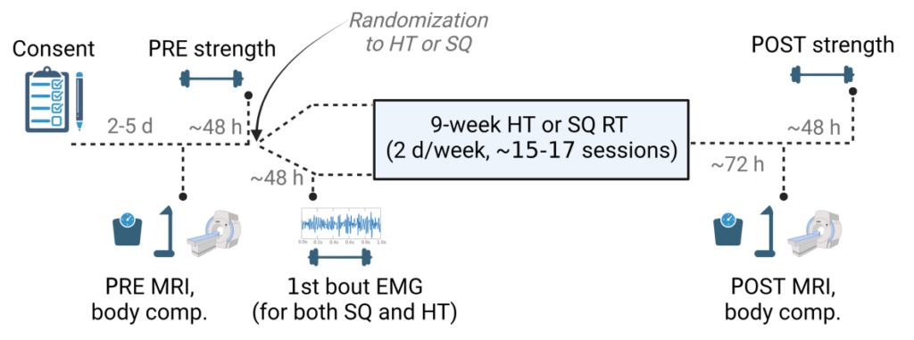
Abbreviations: PRE, pre-intervention testing visit; 140 POST, post-intervention testing visit; HT, barbell hip thrust; SQ, barbell squat; body comp., body composition testing 141 using bioelectrical impedance spectroscopy; MRI, magnetic resonance imaging; sEMG, electromyography.
Evaluation of body composition and MRI measurements
Before testing, participants were instructed to fast for 8 hours, avoid alcohol for 24 hours, refrain from intense exercise for 24 hours, and ensure proper hydration. Upon arrival, participants provided a urine sample for urine-specific gravity assessment, which indicated sufficient hydration (USG levels ≤ 1.020). Heights were measured using a stadiometer, and body mass was determined using a calibrated scale. Body composition was assessed through bioelectrical impedance spectroscopy using a 4-lead SOZO device, providing measurements of fat-free mass, skeletal muscle mass, and fat mass.
For MRI assessment, gluteus maximus muscle cross-sectional area (mCSA) was measured. Participants were positioned in a prone position on the patient table of the MRI scanner at the Auburn University MRI Research Center. After a brief delay, a T1-weighted turbo spin echo pulse sequence was utilized to obtain transverse image sets. The scans consisted of 71 slices with a thickness of 4 mm and no gap between slices. All measurements were conducted by the same investigator (R.J.B.), who was blinded to the participants’ training conditions.
Evaluation of strength levels
Participants performed a wall push test to measure isometric muscle strength. Using a tri-axial force plate, horizontal force production in newtons (N) was recorded. The test involved pushing against a wall with the dominant leg while maintaining a standardized posture. Two repetitions of three seconds each were performed, and the highest peak horizontal force was analyzed.
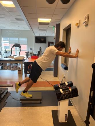
Figure 2. depicts the wall push test with one of the co-authors (M.D.R.)
Dynamic lower body strength was assessed through 3-repetition maximum (3RM) testing for the barbell back squat, barbell hip thrust, and barbell deadlift exercises. Warm-up sets were performed to gauge movement proficiency, followed by sets of three repetitions with incremental load increases. Each exercise was performed with controlled movements, specific depth or apparatus settings, and proper posture. Successful lifts required full knee and hip extensions.
Electromyographic (EMG) recordings during the initial training session
Subjects were instructed to wear loose athletic clothing for the placement of EMG electrodes. Prior to electrode placement, any excess hair was removed, and the skin was cleaned with an alcohol swab. Wireless sEMG electrodes were attached to the right upper gluteus maximus, mid gluteus maximus, lower gluteus maximus, and gluteus medius. Specific placement locations were determined based on established recommendations. A quality check was conducted to ensure signal validity. Maximum voluntary isometric contractions (MVIC) were performed before 10RM testing. EMG signals were recorded during the 10RM sets of the barbell back squat and barbell hip thrust exercises. Signal processing and analysis were performed using dedicated software. EMG data with artifacts were excluded from the analysis.
Procedures for resistance training
The resistance training (RT) protocol involved 3–6 sets per session of either barbell hip thrusts for HT participants or barbell back squats for SQ participants. Except for the first week with one session, the following 9 weeks included two sessions per week on non-consecutive days. The set schemes progressed as follows: week 1, 3 sets; week 2, 4 sets; weeks 3–6, 5 sets; weeks 7–9, 6 sets. Participants aimed for a repetition range of 8–12, adjusting the load accordingly if they fell below or exceeded this range. D.L.P. and 1–2 other co-authors supervised all sessions and motivated participants to reach volitional muscular failure, where they couldn’t perform another concentric repetition while maintaining proper form. The exercise form and tempo matched those described in the strength testing section, with squats allowing the lowest achievable depth. Participants were advised to avoid additional lower-body resistance training outside the supervised sessions, and missing up to 2 sessions was acceptable for analysis.
Data analysis and figure creation
The data underwent analysis using Jamovi v2.3 (https://www.jamovi.org) and R (version 4.3.0). Three sets of analyses ere conducted. Initially, researchers compared the average and maximum sEMG amplitudes of HT and SQ during the initial training session using paired t-tests. Secondly, they examined the longitudinal impact of HT and SQ training on mCSA and strength. Lastly, they explored the predictive value of sEMG amplitudes from the first session on subsequent growth.
Results
Figure 3 displays the CONSORT diagram. A total of 18 participants in the HT group and 16 participants in the SQ group completed the study and were included in the data analysis, except for cases where technical issues prevented data inclusion (e.g., sEMG clipping). The baseline characteristics of the 18 HT participants who completed the intervention were as follows: age: 22 ± 3 years old, BMI: 24 ± 3 kg/m², consisting of 5 males and 13 females. The baseline characteristics of the 16 SQ participants who completed the intervention were as follows: age: 24 ± 4 years old, BMI: 23 ± 3 kg/m², consisting of 6 males and 10 females. Notably, HT participants missed an average of 0.8 ± 0.4 workouts during the study, while SQ participants missed 0.8 ± 0.5 workouts.
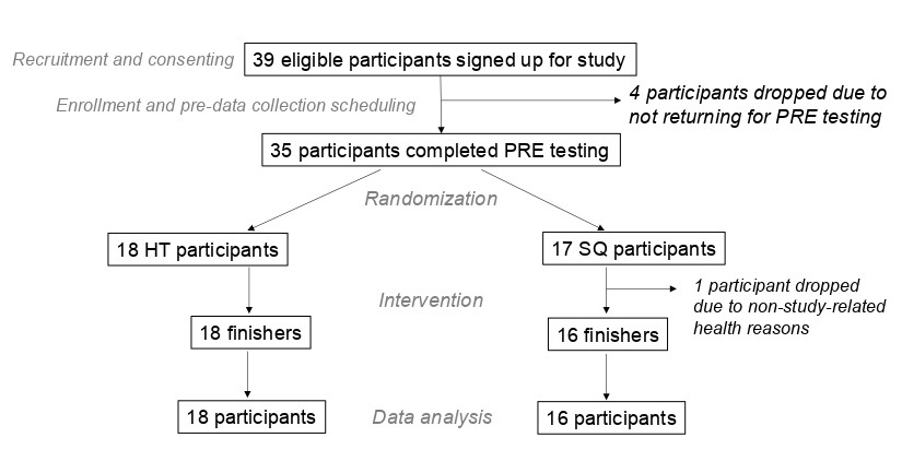
Figure 3. depicts participant numbers through various stages of the intervention. All participants were included in data analysis unless there were technical issues precluding the inclusion of data (e.g., EMG clipping).
Figure 4. presents the sEMG results of the initial workout, specifically the data obtained from the right gluteus muscles during a single set of 10RM hip thrust and a single set of 10RM squat. The mean sEMG values were significantly higher during the hip thrust compared to the squat set (p < 0.01 for all sites; Fig. 4b). The peak sEMG values were significantly greater for the upper and middle gluteus maximus (p < 0.001 and p = 0.015, respectively), with only minor differences observed for the lower gluteus maximus or gluteus medius sites (Fig. 4b). The number of repetitions completed during the 10RM sets used for sEMG recordings did not differ between the two exercises (back squat: 9±1 repetitions, hip thrust: 9±2 repetitions).
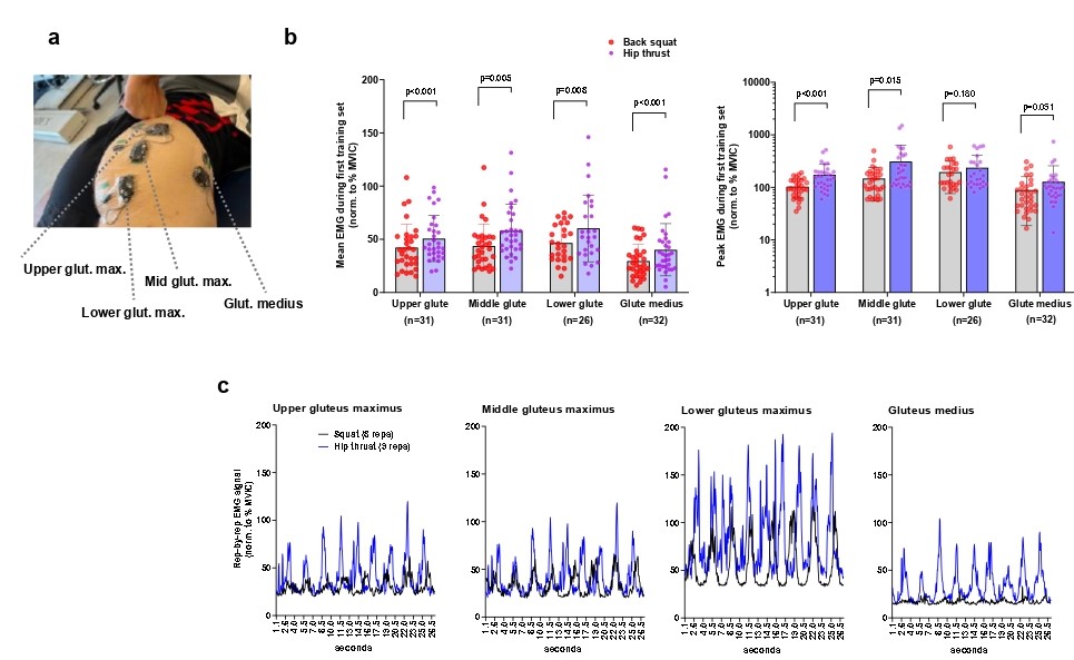
Legend: During the first session, all participants performed both back squats and barbell hip thrusts while we recorded sEMG amplitudes. (a) Representative sEMG electrode placement is depicted on a co-author in panel. (b) Data depict mean (left) and peak (right) sEMG amplitudes during one 10RM set of hip thrusts and one 10RM set of back squats. As 34 participants partook in this test, sample sizes vary due to incomplete data from electrode slippage or clipping. Bars are mean ± SD, and individual participant values are depicted as dots. (c) Representative data from one participant.
The impact of back squats (SQ) compared to hip thrusts (HT) on the combined left and right muscle cross-sectional area (mCSA) of the gluteus muscles was minimal. The estimated effects slightly favored HT for the lower [-1.6 ± 2.1 cm²; CI95% (-6.1, 2.0)], mid [-0.5 ± 1.7 cm²; CI95% (-4.0, 2.6)], and upper [-0.5 ± 2.6 cm²; CI95% (-5.8, 4.1)] gluteal mCSAs, although these estimates were overshadowed by the variability. The combined mCSA values for the gluteus medius + minimus showed a smaller degree of growth (refer to Table 1), with an estimated effect that also slightly favored HT but exhibited noticeable variability [-1.8 ± 1.5 cm²; CI95% (-4.6, 1.4)].
Legend: Figure depicts change adjusted for pre-intervention scores for MRI-derived muscle cross-sectional area (mCSA). (a) left + right (L+R) upper gluteus maximus, (b) L+R middle gluteus maximus, (c) L+R lower gluteus maximus, and (d) L+R gluteus medius+minimus. Data include 18 participants in the hip thrust group and 16 participants in the back squat group. Graphs contain change scores with individual participant values depicted as dots.(e) Three pre and post representative MRI images are presented from the same participant with white polygon tracings of the L+R upper gluteus maximus and gluteus medius+minimus (top), L+R middle gluteus maximus (middle), and L+R lower gluteus maximus (bottom).
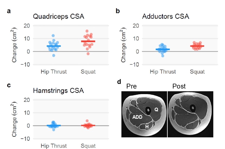
When comparing to hip thrusts (HT), back squats (SQ) resulted in more substantial growth in muscle cross-sectional area (mCSA) for the quadriceps [3.6 ± 1.5 cm²; CI95% (0.7, 6.4)] and adductors [2.5 ± 0.7 cm²; CI95% (1.2, 3.9)]. However, the growth of hamstrings showed no significant differences between the two conditions, with minimal effects observed [0.1 ± 0.6 cm²; CI95% (-0.9, 1.4)].
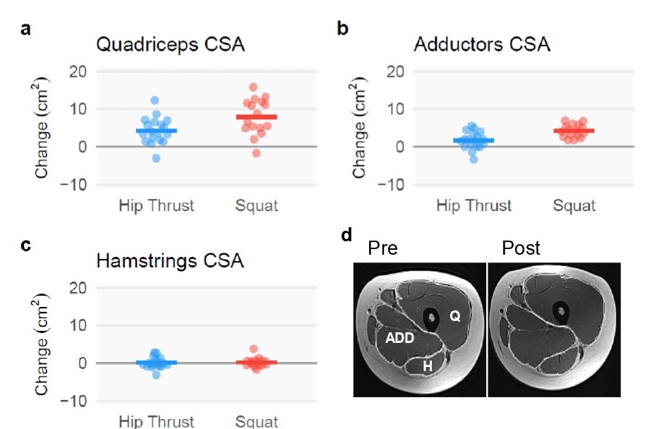
Legend: Figure depicts change adjusted for pre-intervention scores for MRI-derived muscle cross-sectional area (mCSA). Left and/or right (a) quadriceps, (b) adductors, and (c) hamstrings. Data include 18 participants in the hip thrust group and 16 participants in the back squat group. Bar graphs contain change scores with individual participant values depicted as dots. (d) A representative pre- and post-intervention MRI image is presented with white polygon tracings of the quadriceps (denoted as Q), adductors (denoted as ADD), and hamstrings (denoted as H).
Regarding strength outcomes, the specific lift 3RM values indicated a preference for the respective group allocations between back squats (SQ) and hip thrusts (HT). Specifically, the 3RM value for squats favored SQ [14 ± 2 kg; CI95% (9, 18)], while the 3RM value for hip thrusts favored HT [-26 ± 5 kg; CI95% (-34, -16)]. The results were less definitive for the deadlift 3RM [0 ± 2 kg; CI95% (-4, 3)] and wall push [−7 ± 12 N; CI95% (-32, 17)].

Legend: Figure depicts change adjusted for pre-intervention scores for (a) 3RM barbell back squat values, (b) 3RM barbell hip thrust values, (c) 3RM barbell deadlift values, and (d) wall push as demonstrated in Figure 2. Data include 18 participants in the hip thrust group and 16 participants in the back squat group.
Discussion
To explore hip extensor exercises and the validity of theory and acute measures for exercise selection, researchers compared back squats and barbell hip thrusts. Acutely, HT showed higher sEMG amplitudes, but this didn’t translate to longitudinal adaptations. Modest differences in gluteus muscle hypertrophy were observed, except for the gluteus medius and minimus. SQ favored adductors and quadriceps hypertrophy, while hamstrings showed no significant growth in either group. Strength outcomes favored HT for hip thrusts and SQ for back squats. sEMG amplitudes were unreliable predictors of hypertrophy. They discussed these results and their implications for exercise prescription.
Hypertrophy Outcomes
Similar gluteus maximus hypertrophy found in upper, middle, and lower regions after 9 weeks of squat or hip thrust training, despite evidence favoring tension-induced growth in lengthened positions. Prior research focusing on isolated muscle work raises questions about the impact of loading context on adaptations. Muscle contributions, and not just positions, may need to be jointly considered in determining whether superior hypertrophy outcomes would be achieved. sEMG and musculoskeletal modeling imply less gluteus maximus recruitment at the bottom of the squat, hindering stretch-induced hypertrophy. Motor control and study-specific factors also influence gluteus maximus contribution and adaptation. Both exercises may lead to comparable muscle hypertrophy in untrained individuals due to the rapid early growth effects of resistance training. Novice trainees may experience comparable gluteal muscle hypertrophy with a nine-week training program involving either the hip thrust or squat. Thigh hypertrophy is greater with the squat, aligning with previous findings. The adductor magnus is prominently recruited during the squat, particularly at the bottom, suggesting its contribution to the exercise. Adductor magnus mCSA changes correlate with increased squat depth. Hamstring mCSA remained unchanged in both groups, consistent with prior research. These findings suggest that the hip thrust predominantly focuses on gluteal muscle hypertrophy while minimizing growth in non-gluteal thigh muscles. In essence, the hip thrust seems to specifically target the gluteus Maximus.
Strength Outcomes
Both groups saw significant improvements in strength across all tested exercises. However, hip thrust resistance training (HT RT) showed greater enhancements in hip thrust strength, while back squat resistance training (SQ RT) resulted in higher back squat strength gains, which aligns with the principle of training specificity. The HT group experienced a 63% increase in hip thrust strength and a 17% increase in back squat 3RM, whereas the SQ group demonstrated a 34% increase in hip thrust strength and a 44% increase in back squat 3RM. Interestingly, both groups exhibited similar improvements in deadlift and wall push outcomes, with a 15% increase in deadlift strength for the SQ group and a 16% increase for the HT group, along with a 7.6% increase in wall push strength for the SQ group and a 10% increase for the HT group.
Conclusion
Both squat and hip thrust resistance training resulted in comparable gluteal hypertrophy, but squat training produced greater hypertrophy in the quadriceps and adductors. Additionally, both forms of resistance training showed similar strength improvements in the deadlift and wall push exercises. Notably, these outcomes couldn’t be accurately predicted based on acute data (sEMG). These findings offer valuable guidance to trainees regarding two popular hip-specific exercise methods, enabling them to make informed exercise choices based on their specific structural or functional objectives.
Limitations
This study has several limitations to consider. Participants were young untrained individuals, so generalization to other populations may be limited. The study duration was short, and the focus was on gluteal hypertrophy. Compression from the MRI coil may have affected thigh musculature, and distal measures were not obtained. The training protocol involved equated volume and two training days per week, limiting generalizability. Menstrual cycle phase and contraceptive usage were not considered, but evidence suggests they have minimal impact on muscle hypertrophy and strength gains during resistance training. While acknowledging that a sample size of 34 participants is typical for resistance training research, it would be beneficial to include larger groups, particularly consisting of trained individuals.
To access the original study click here.
Reviewed and prepared by:
Željko Banićević, Ph.D.*, M.Sc., B.Sc.

